The brain
The brain is one of the most fascinating organs in the human body. It not only enables us to interact with our environment by integrating the information we gain through our senses, but also allows for creativity and the creation of things which have not existed before.
Anatomy
In particular in the early stages during development, the brain undergoes significant changes. The brain is considered to form when the neural plate closes and becomes the neural tube at around 3 weeks of post-menstrual age (PMA). After this point, three principal enlargements form, namely the forebrain (proencephalon), the midbrain (mesencephalon) and the hindbrain (rhombencephalon). Each of these enlargements will form an important part of what is called the adult human brain. The individual parts of the brain and what they develop into is shown in the next figure.
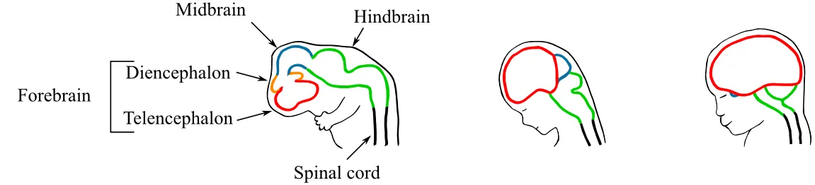
As we can see, the forebrain is split up into two parts, the diencephalon, which is the basis, for example, for our eyes, and the telencephalon, which forms the largest part of the human brain, the cerebral hemispheres. It is in the telencephalon, where most of our higher cognitive functions take place.
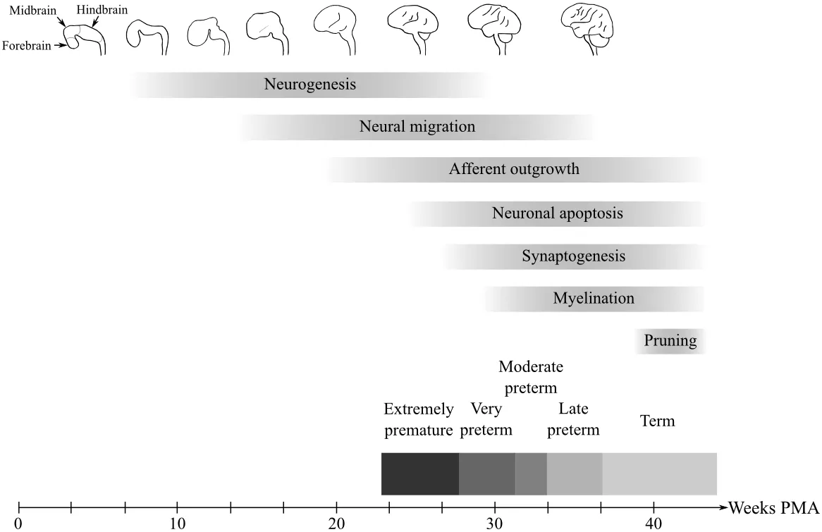
So far we have talked about what scientists call grey matter, the set of neural cells which are responsible for processing information. The name "grey matter" stems from the colour of the cell bodies. However, in order for the individual cells to communicate, white matter is necessary. These connections appear white due to the fatty material (myelin) that is used to insulate them and which increases their efficacy with respect to information transport. The development of both grey and white matter are described in more detail below. The following figure summarises the key parts of brain development during pregnancy.
Sometimes babies are born too early (premature). One important question is how this event influences the development of the baby's brain. Prematurity and its effects are also described below.
Gray matter
Grey matter refers to the part of the brain that is responsible for the processing of information. It integrates the information which we acquire through our senses and transforms them into actions, which are then executed by our bodies. In general we can distinguish between two scales at which this development takes place, the microscopic scale (development of cells) and the macroscopic scale, defined by the development of valleys (sulci) and hills (gyri) in a process called cortical folding.
Microscopic
One of the most important time frames during microscopic grey matter development is the period between 24 and 40 weeks. The major events which take place in this period are called neurogenesis, neural migration and neural apoptosis.
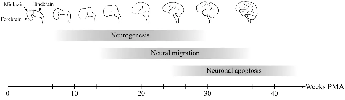
Neurogenesis refers to the creation of neurons. It mainly occurs during the second trimester. In most parts of the brain no new neurons are created after birth. One important exception is the hippocampus, where the neurons are linked with memory formation. However, from 24 weeks of gestation until around 4 weeks after birth, rapid cell death occurs as well, reducing the total number of neurons by half.
Neurons do not stay at the place where they are created. After they are produced, they migrate towards the developing neocortex. They do so by using a scaffolding (basal processes) to which they attach themselves. In particular, the first migrating neurons form a structure known as preplate. This structure splits then into two layers (marginal zone and subplate), between which the cortical plate forms.
Macroscopic
At around 3 weeks the neural tube, the ancestor of the brain, forms. From there three principal enlargements form, the forebrain (proencephalon), the midbrain (mesencephalon) and the hindbrain (rhombencephalon).
Over the course of pregnancy, the hindbrain develops into the cerebellum, pons and medulla oblongata, whereas the proencephalon first splits into the diencephalon and the telencephalon. The diencephalon continues to develop into the optic vesicles (the retina of eyes), the thalamus (responsible e.g. for distributing information) and the hypothalamus (responsible e.g. for regulating temperature or hunger). The telencephalon, on the other hand, forms the largest part of the human brain, the cortex.
The cortex starts to form sulci and gyri in a process called cortical folding from approximately 15 weeks of gestation onwards. Sulci ("valleys") and gyri ("hills") form the major landmarks of the human brain. The primary sulci are already present by 28 weeks, however smaller, secondary and tertiary sulci form afterwards and continue to form after birth.
White matter
White matter refers to the structural connectivity between neurons, the processing units of the brain. After neurons migrate to their destination they develop outgrowths, called axons. These outgrowths can be seen as the pipes within the brain that allows the processing units to talk to each other. In order to do so, neurons use electro-chemical signals to transfer information along the axons. Here we will introduce the concepts of synaptogenesis, myelination and pruning.
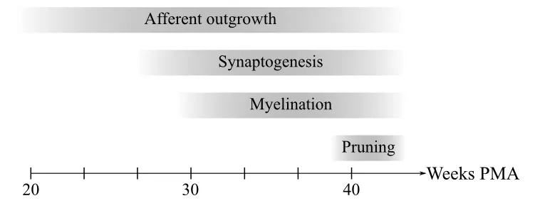
Around 24-26 weeks post-menstrual age (PMA) the first axons (the pipes) grow towards the cortical plate. Once they reach their target, they connect to the neurons through synapses in a process called synaptogenesis. These connections allow for the differentiation of the different parts of the cortex that govern for example movement and sensation. Axons continue to grow until shortly after birth, however, synapses are being formed until 2 years of age.
One important aspect of synaptogenesis is that the brain "over-connects" its regions in the beginning. This means that more synapses are formed than are necessary over a lifetime. Therefore, even though the amount of synapses increases rapidly after birth for a short period of time, a process called pruning starts to take over, which reduces their total numbers. During development different parts of the brain reach their maximum synaptic density at different time points, however, in general we lose approximately 40% over our lifetime.
Communication within the brain occurs through electro-chemical signals. One aspect of this kind of information transport is that it loses signal intensity the longer the information has to travel. The brain counters this loss by using myelin sheathes as insulation. Around 29 weeks PMA, myelin sheathes start to form around axons, which allow for much better signal transfer. Myelin is made of a fatty substance with a white colour, which is the reason why the system of axons or structural connectivity is referred to as white matter. The process of myelination continues even into late adolescence.
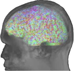
One way to investigate the structural connections is by using diffusion magnetic resonance imaging (dMRI). With dMRI researchers can estimate where bundles of axons are and which brain regions connect with each other.
Vasculature of the brain
As the brain is the organ with the highest "maintanance cost" in our body, it becomes imperative that its supply lines, or blood vessels, are adequate. However, considering how complex the brain is, the system of blood vessels, i.e., the vasculature, can be considered equally complex. Through the vasculature, the brain is supplied with oxygen, as well as nutrients, which make its daily work possible.
Blood is supplied to the head through two major sets of arteries, the left and right common carotid artery, as well as the left and right vertebral arteries. Part of the carotid artery system, the external cartoid arteries, can easily be identified, when trying to take your pulse at the neck using your fingers. However, the brain's blood supply is handled by the vertebral arteries, and the internal carotid arteries. Within the skull, these arteries combine to form the Circle of Willis.
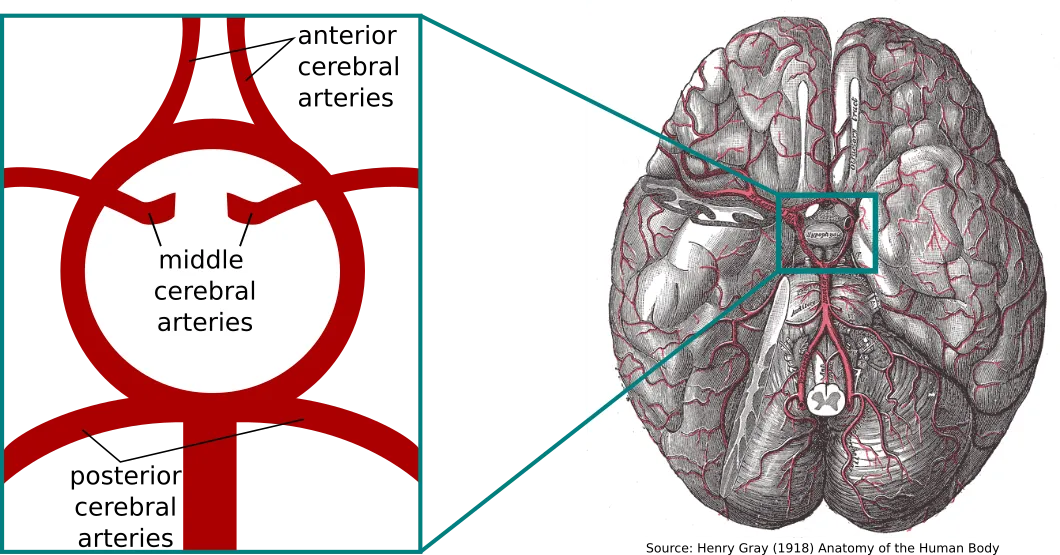
From the Circle of Willis, three major arteries branch off which supply the cortex. The anterior cerebral artery extends upwards and supplies the frontal lobes (towards the forehead). These areas are assumed to support logical thought, personality, and voluntary movement. The middle cerebral artery is the largest branch and supplies the biggest part of the brain (towards the sides of the head). It also supplies part of the frontal lobe, as well as the temporal and parietal lobes of the brain. These areas include primary motor and sensory areas of the face, arms and hands, as well as speech. The third artery is the posterior cerebral artery (towards the back of the head). It supplies part of the temporal lobes, as well as the occipital lobe, which is repsonsible for visual processing.
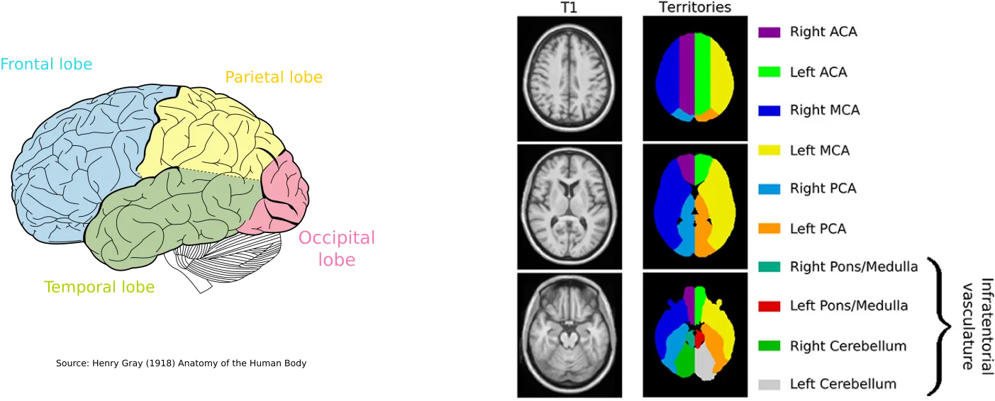
We can subdivide the cortex of the brain based on their supplying arteries. These areas are commonly referred to as vascular territories. While the supratentorial part of the brain (the region above the cerebellum), can be differentiated based on these arteries, the infratentorial part can be divided based on anatomical landmarks. The figure above shows where the vascular territories are located within the brain based on a template we created as part of our studies.
In addition to these large, major arteries, many more smaller arteries are distributed throughout the brain to support, e.g., the white matter. The microvasculature of the brain is particularly hard to study, as it requires high-resolution imaging techniques. While significant progress has been made in imaging small vessels in the human body in-vivo by means of new, more powerful magnetic resonance imaging, or super-resolution ultrasound, they each have limitations in terms of acquisition time or loss of signal due to the properties of the skull. Therefore, the most prevalent method for characterization still remains post-mortem histological approaches. However, how much the vasculature at this scale changes after death, is still an open question, meaning there is still a lot for us to learn about the brain's microvasculature.
Medical Conditions and Diseases
Prematurity
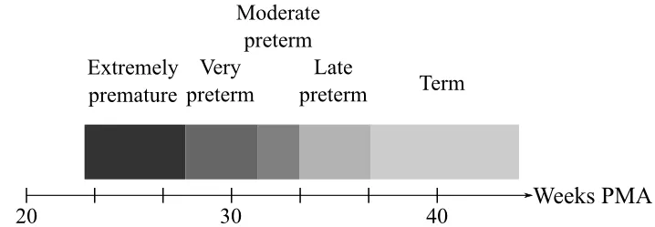
Sometimes babies are born premature, which means before 37 weeks of pregnancy. Based on their post-menstrual age at birth, premature babies can be divided into extremely preterm (before 28 weeks), very preterm (between 28-31 weeks), moderate (32-33 weeks) and near term or late preterm (34-37 weeks).
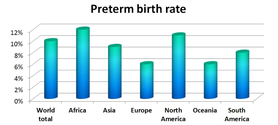
Although the causes for premature birth are not completely understood, prematurity is not uncommon. A combination of multiple factors may play a role, such as socio-economic factors, genetic influences, medical conditions, pregnancy history or due to the use of assisted reproductive technologies. The World Health Organization (WHO) has estimated that premature birth occurs in one out of ten pregnancies world-wide. It has also been indicated that the amount of premature births is increasing.
However, with advancements in perinatal care, the number of prematurely born babies that survive is increasing as well, which can result in high physical, psychological and economical costs.
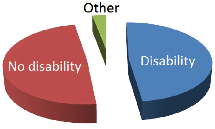
Preterm babies undergo substantially different brain development, compared to term-born babies. Unfortunately this can lead to neurodevelopmental impairment. Such impairments can include motor, auditory, visual and cognitive function or mental disorders such as autism or attention deficit hyperactivity disorder (ADHD). Wood and colleagues, for example, have suggested that approximately 50% of extremely preterm infants have some sort of impairment at 30 months of age.
Unfortunately, the question of how to determine which premature babies need help in the long run remains an open challenge. Therefore, the investigation of neonates (new born babies) is of great interest with much effort expended on finding ways of identifying the group of neonatal patients that need special attention. Special attention in this case means the possibility of an early intervention to either reduce severity of the condition or prevent negative long-term outcomes. Furthermore, targeted support is also of great importance, not only for the children themselves, but also for their families.
Stroke
In simple terms, stroke can be seen as a distrubance in blood flow to the brain. Stroke was first recognized by Hippocrates over 2400 years ago. Back then it was referred to as apoplexy, which roughly translates to "struck down by violence". Even these days, we can identify strokes as sudden appearance of symptoms, such as paralysis.
To identify a person with stroke, one can utilize the acronym F A S T. Each letter stands for a commonly observed symptom, ending with a recommendation to go to a hospital:
- Facial drooping - A section of the face is affected and drooping and hard to move, resulting in a crooked smile.
- Arm weakness - Not being able to raise both arms fully horizontally.
- Speech difficulties - Speaking or understanding speach is difficult or impossible.
- Time: If any of these symptoms occur, go to the hospital immediately. Time is of the essence, as full treatment may only be possible in the early stages.
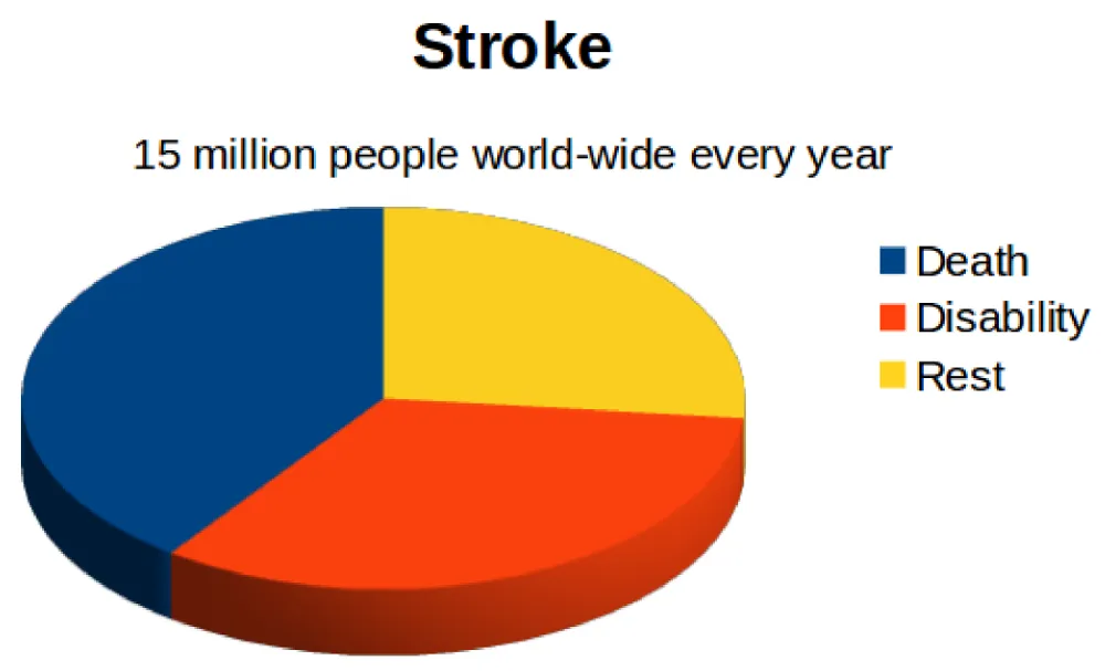
Stroke has been part of humans for millenia. However, stroke is not a rare occurance. Every year, 15 million people world-wide are reported to have a stroke, where about 75% of patients either die, or end up with a disability. But not all strokes are equal. Generally, we differentiate between haemorrhagic and ischemic strokes. Out of all strokes, about 15% are haemorrhagic, whereas 85% are ischemic.
Haemorrhagic strokes are a result of a ruptured blood vessel, leading to a bleed within the brain. Due to the accumulating blood, the surrounding brain tissue gets compressed, which can severly damage important brain areals.
Ischemic strokes, on the other hand, are caused by a blockage of a blood vessel, which distrubes the blood supply to regions within the brain. While other vessels may help to compensate some of the lacking blood supply, the longer this ischemia takes place, the more of the under-/unsupplied brain regions will become necrotic and die off.
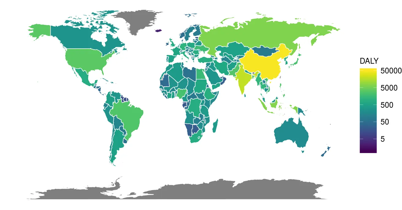
Stroke is a prevalent disease, which occurs all over the world. However, it is difficult to assess the impact that stroke has on a populations. Some patients with strokes will die, while others may become disabled. A concept which has been widely used aiming to summarize both is the so-called disability-adjusted life year (DALY). In short, it can be considered as loss of years of "healthy" life. It does so by summing up the years of life lost and the years lost due to disability. The following figure shows the DALY throughout the world in case of stroke.
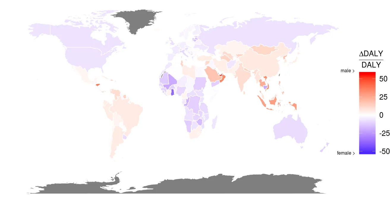
It is important to acknowledge that stroke does not affect everyone in the same manner. There has been a well reported differnce in strokes between men and women, where women usually have stroke later in life. However, these strokes are often more severe compared to men. While this is still an open research question, it is also important to acknowledge that there might be a survival bias, as men, on average, die at a younger age. The following figure illustrates the difference in percent points of DALY comparing men and women with respect to the DALY within each country. Red colors indicate that men account for more of the total DALY in the country, whereas blue colors indicate that stroke in women account for more of the total DALY.
Sex is not the only aspect that results in different stroke prevalence and outcome. In general, we can differentiate between two types of factors: predetermined and modifiable. Predetermined factors include for exmaple family history of stroke, genetics, and age. While we cannot alter these aspects, knowing and understanding them can help us develop theraputic and targeted treatments. In contrast, modifiable factors can significantly reduce the risk of suffering from a stroke. These include for example physical activity, healthy lifestyle, diet, alcohol consume, as well as smoking. Importantly, one relatively recent study suggested that bad lifestyle and good genetics can be just as detrimental as bad genetics with good lifestyle.